Our main research interests
The Amberg group at the Medical University of Vienna focuses on three main topics:
- Deciphering the molecular and cellular mechanisms of faithful human brain development
- Identifying human-specific aspects of brainstem development
- Modeling developmental diseases of the human brain in vitro using patient-derived iPSCs and regionally patterned organoids, studying:
- pediatric brain tumors from individuals presenting with ultra rare tumor predisposition syndromes
- GNB1E (GNB1 encephalopathy), an ultra-rare neurological disorder
Our research involves mouse models, patient-derived iPSCs, forebrain and brainstem organoids, and most importantly human brain tissue derived from my Division’s resourceful neurobiobank.
The biobank harbors a developmental brain bank with fetal brain tissue across developmental stages starting from GW14 (controls and neurodevelopmental malformations), as well as pediatric, adult and aged brain, brain tumors, tissue affected by epilepsy, neuroinflammatory or neurodegenerative diseases, muscle biopsies, nerve biopsies, gut biopsies, serum and CSF. Feel free to reach out for access to our neurobiobank, we are happy to collaborate.
Selected current projects:
- Science Communication - FWF Project SCP7695224
- Research - FWF Project V1041
- Research - WWTF Project LS25-014
- Research - soon to be started FWF Project "BrainStem"
Our past and current funding:
Bridging Clinical and Fundamental Neuroscience
Being positioned in the clinical setting of the Division of Neuropathology and Neurochemistry at the Medical University of Vienna grants us direct access to precious patient material and excellent state-of-the-art expertise in neuropathology and molecular diagnostics. Thus, we are generating iPSCs from patients presenting with tumor predisposition syndromes as well as GNB1 encephalopathy, and cultivate brain organoids with distinct regional identity to study human disease in vitro.
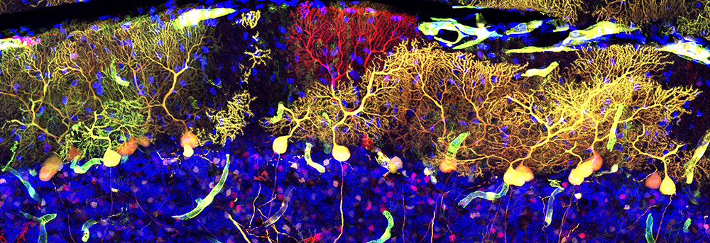
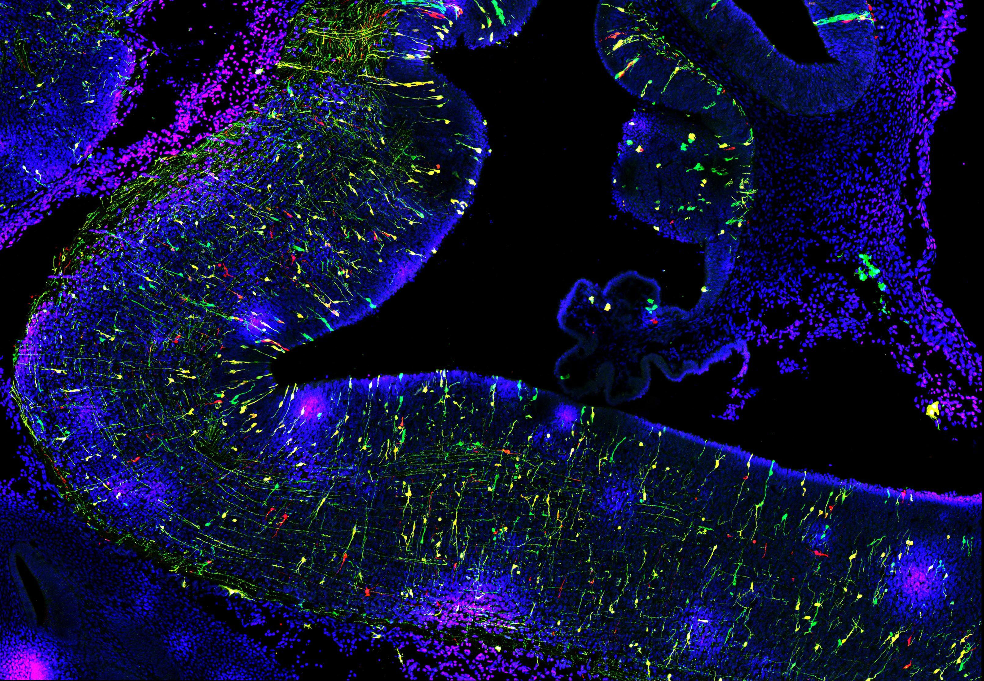
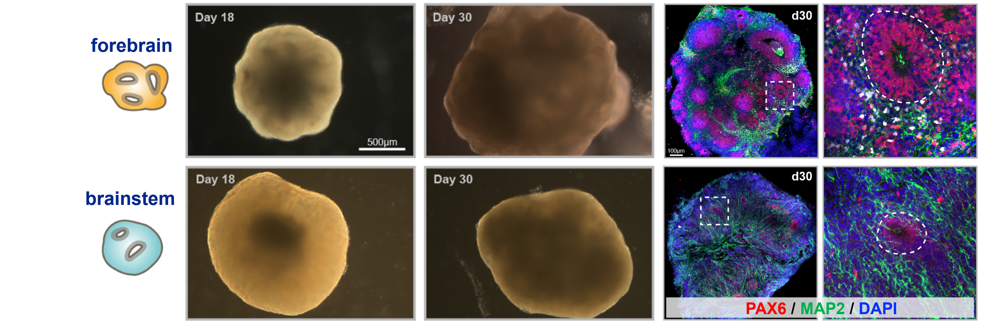
Examples of our regionally patterned cerebral organoids:
News
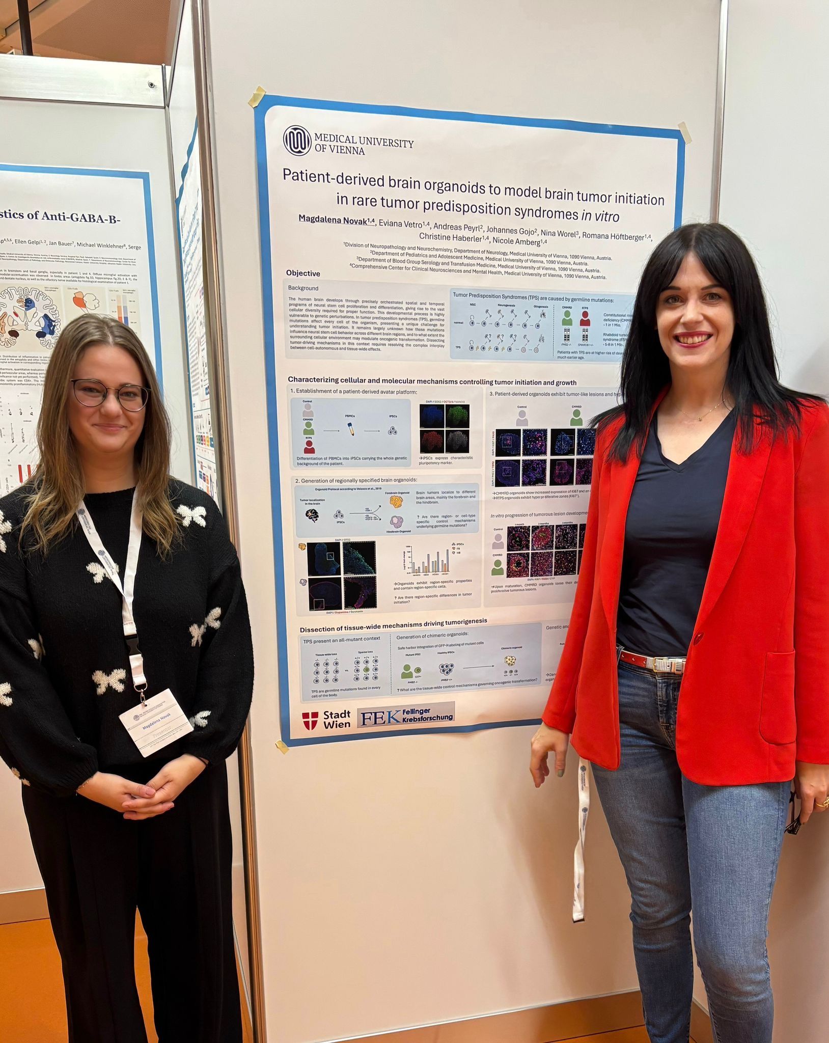
Best Poster Award at YSA Symposium 2026 for Magdalena Novak
Magdalena is a highly talented Master's student in the Master program Neuroscience at the University of Vienna. She is reprogramming patient-derived PBMCs and has established regionally specified brain organoids (forebrain and brainstem) from our patient-derived iPSCs.
She has been able to show that patient-derived forebrain organoids develop hyperproliferative zones and tumor-like lesions, while at the same time displaying a dysintegration of rosettes. During her thesis, she was able to assess whether brain organoids with distinct regional patterning show different dynamics in tumor initiation and has applied distinct transcriptomics approaches to unravel the molecular profile of her patient-derived organoids.
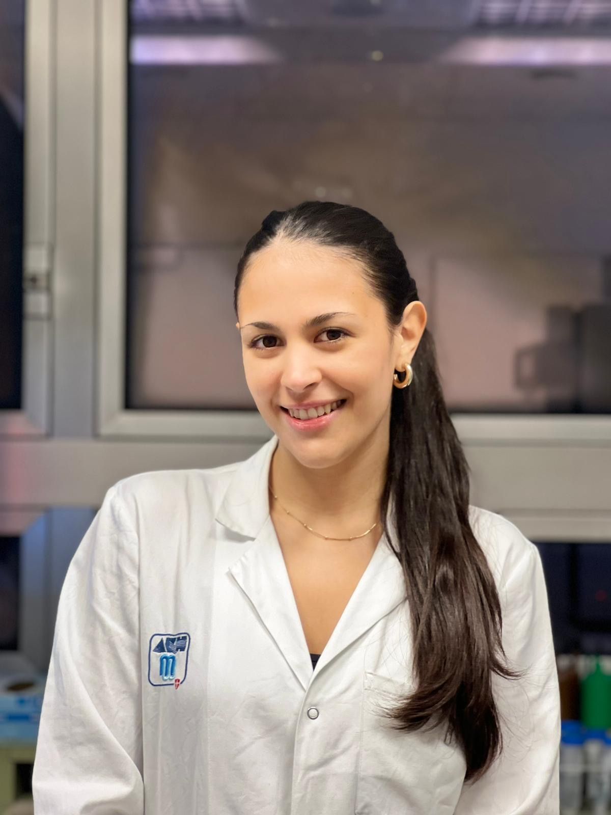
Alumna: Eviana Vetro (erasmus+ student)
Eviana is a Master's student from the University of Cologne, who performed pluripotency validation in one of our patient-derived iPSC lines. She could also show comparable behavior of healthy donor-derived iPSCs and patient-derived iPSCs in terms of proliferation rate, expression of pluripotency markers and ability to give rise to cerebral organoids.
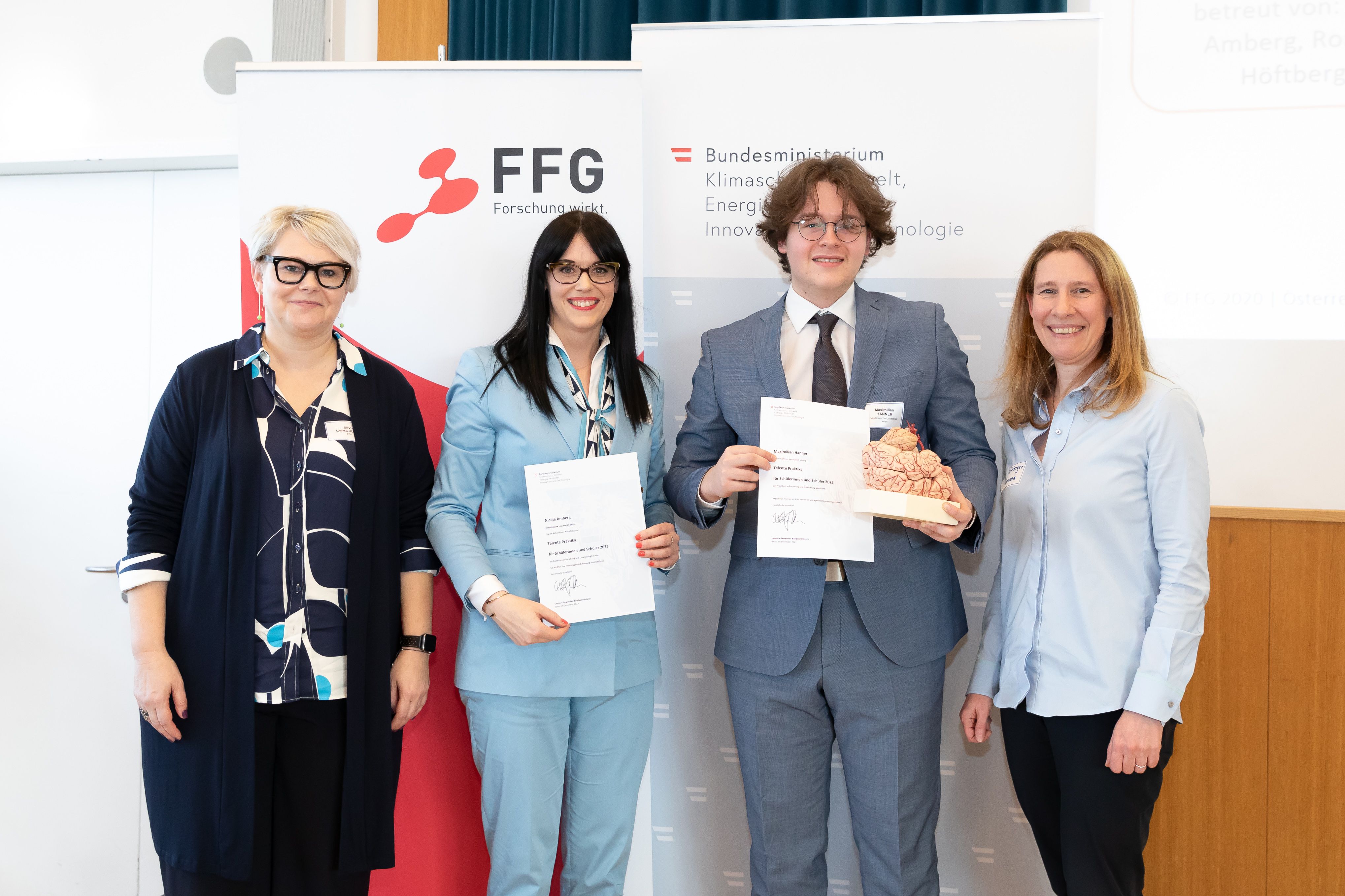
FFG Research Award for Alumnus Maximilian Hanner (FFG intern)
Maximilan attends Med School at the Medical University of Vienna. He has performed histological stainings for H3K27me3 on human fetal cerebellar tissue, where he observed dynamic, cell type-specific changes in H3K27me3 levels in the course of cerebellar development. He also became interested in a rare cerebellar malformation called rhomboencephalosynapsis, which shows distinct H3K27me3 levels.